
 (NABH Accredited)
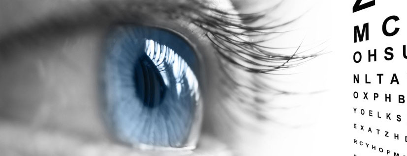

- CATARACT
- LASERS
- REFRACTIVE SURGERY
- CORNEAL SERVICES
- GLAUCOMA
- MEDICAL RETINA
Glaucoma is a group of conditions characterised by a progressive loss of vision. Glaucoma is classified as primary or secondary. The most common type of glaucoma, called "primary" open angle glaucoma, is a chronic disturbance of the normal fluid pressure inside the eye and is generally age-related. "Acute" or secondary closed angle glaucoma is more rare and is identified by a sudden, painful shutting down of the mechanisms controlling intraocular fluid pressure. In "secondary" glaucoma, factors such as trauma, certain drugs, infections, tumours or advanced cataracts cause an increase in the intraocular fluid pressure.
Glaucoma is the second leading cause of blindness worldwide. It is a heterogeneous group of disorders marked by damage to the structural or functional integrity of the optic nerve that causes characteristic atrophic changes. Over time, this may also lead to specific visual field defects. Damage can be arrested or diminished by adequate lowering of intraocular pressure (IOP). Yet, some debate still exists as to whether IOP should be included in the definition of glaucoma, as some subsets of patients can exhibit the characteristic optic nerve damage and visual field defects while having an IOP within the normal range. PRESSURES DO NOT DETERMINE IF YOU HAVE GLAUCOMA. You can have normal or low pressure and have glaucoma. It is thought up to 50% of people with glaucoma have normal tension glaucoma. Your eye doctor must examine your optic nerve head to diagnose glaucoma.
The generic term “glaucoma” refers to the entire group of glaucomatous disorders as a whole, because multiple subsets of glaucomatous disease exist. Glaucoma is not just a disease of IOP but rather a multifactorial optic neuropathy. A more precise term should be used to describe the glaucomatous disorder, if the specific diagnosis is known.
Who is most likely to get glaucoma?
Although anyone can get glaucoma, some people are more at risk than others for "primary" openangle glaucoma. They include: people with a family history of glaucoma, anyone over the age of 60, and African-Americans.
What are the symptoms of glaucoma?
Often glaucoma has no symptoms! Since the conditions are progressive, the earliest symptoms are often mild, such as a slight change in colour vision. Acute glaucoma may initially cause mild bouts of blurred vision, haloes around lights or eye discomfort. However, as the conditions progress, there is eventually a permanent vision loss.
How can glaucoma be detected?
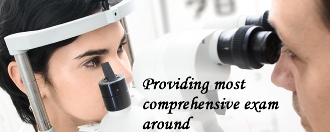
Regular eye examinations are the best hope for early detection, especially for those in the high risk groups. Several tests performed during the eye examination are designed to look for signs of glaucoma. Your eye doctor will review both your general health and your ocular health during your visit as they also provide important clues.
Can glaucoma be treated?
Once detected, glaucoma may respond to drug therapy. If this treatment proves unsuccessful, surgery may be necessary. In the case of secondary glaucoma, the progression of the disease may be stopped by removing the source. Unfortunately, nerve cells do not regenerate once destroyed, therefore any vision loss which has occurred is permanent. Early detection is critical.
Are there precautions a person with glaucoma should take?
The drugs used to treat glaucoma may diminish night vision and peripheral vision. Caution is urged when driving. Also, glaucoma medications may aggravate certain medical conditions, such as emphysema. Certain medications can be potentially harmful when taken along with glaucoma medications. If you are a diabetic or have high blood pressure, your condition or its treatment may affect glaucoma.
DIAGNOSIS OF GLAUCOMA
Besides the basic evaluations done during an eye examination, you may need more specialized tests, depending on your age, medical history and risk of developing eye disease. When evaluating glaucoma patients the doctors at the Sucha Eye Cure Clinic recommend the following:
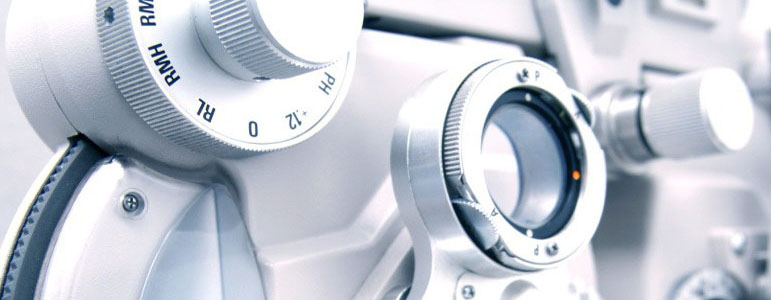
- Pachymetry
This test measures the thickness of your cornea — an important factor in evaluating your intraocular pressure measurement. After applying numbing eye drops, your eye doctor uses an instrument that emits ultrasound waves to measure your corneal thickness. If you have thick corneas, your eye pressure reading may read artificially high even though you may not have glaucoma. Similarly, people with thin corneas can have normal pressure readings and still have glaucoma. The OHTS study concluded that people with thin corneas that were diagnosed with glaucoma had a greater risk of optic nerve damage.
Visual field test (Perimetry 24-2)
Your visual field is the area in front of you that you can see without moving your eyes. The visual field test determines whether you have difficulty seeing in any areas of your peripheral vision — the areas on the side of your visual field. There are a few different types of visual field tests:- Confrontation visual field exam. Your eye doctor sits directly in front of you and asks you to cover one eye. You look directly at your eye doctor while he or she moves his or her hand in and out of your visual field. You tell your doctor when you can see his or her hand or fingers.
- Tangent screen exam. You sit a short distance from a screen and stare at a target at its center. You tell your doctor when you can see an object move into your peripheral vision.
- Automated perimetry. Your eye doctor uses a computer program that flashes small lights as you look into a special instrument. You press a button when you see the lights.
Using your responses to one or more of these tests, your eye doctor determines the fullness of your peripheral vision. If you aren't able to see in certain areas, noting the pattern of your visual field loss may help your eye doctor diagnose your eye condition. :
Optical Coherence Tomography (OCT)- Optical Coherence Tomography is a diagnostic test that provides high-resoulution, cross-sectional imaging of ocular tissues. It is predominantly used for imaging of the back of the eye to measure retinal nerve thickness entering the optic nerve and macular area. OCT is used to study and monitor diseases such as glaucoma and age-related macular degeneration.
- Heidelberg
Retinal Tomograph (HRT3)
This technology is one of the best ways to follow further loss of optic nerve fibers. Glaucoma management is largely about following progression or change in the optic nerve head.